Eyelids
The eyelids cover and protect the outer part of the eye.

UNDERSTANDING YOUR EYES
The precious gift of God, the eye is a wonderful structure with many parts working in unison to provide us with good vision.
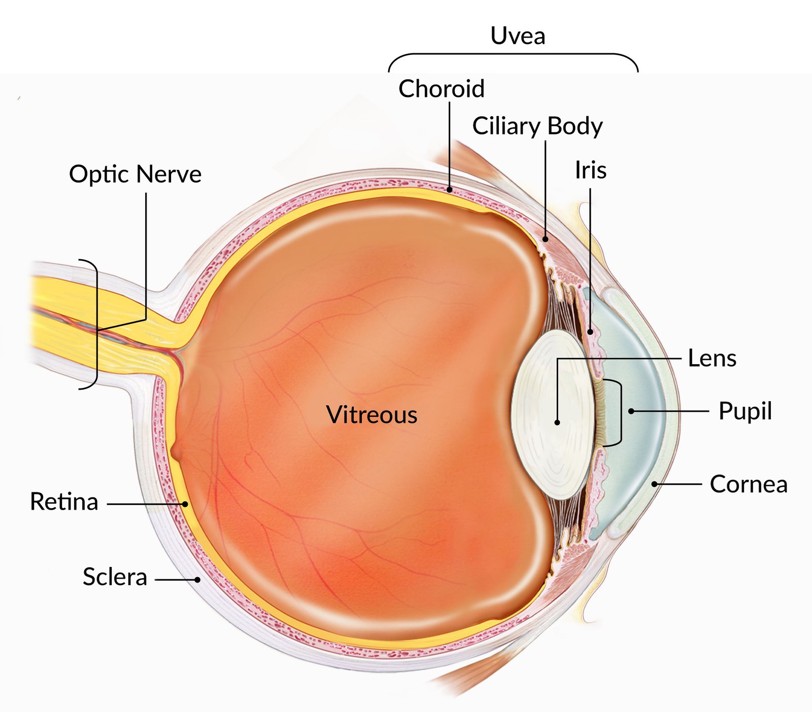
The eye works much like a camera. The cornea and lens focus an inverted image on the retina, the light-sensitive layer at the back of the eye. Signals then travel through the optic nerve to the brain, where they are interpreted as the world we see.
EXPLORE THE EYE
The eyelids cover and protect the outer part of the eye.
The white part of the eye is called the sclera. It is made of tough, fibrous tissue that protects the inner structures of the eye.
The cornea is the transparent window at the front of the eye. It allows light to enter and helps focus it.
Aqueous humour is the clear fluid in the front of the eye, between the cornea and the lens.
The iris is the coloured diaphragm that controls the amount of light entering the eye.
The pupil is the opening in the centre of the iris. The iris adjusts its size in response to light.
The lens is a transparent structure behind the iris. It is held in place by suspensory ligaments, called zonules, and helps focus light on the retina.
Vitreous humour is the clear gel between the lens and the retina. It fills the back of the eye and helps maintain its shape.
The choroid is a pigmented, blood-vessel-rich layer between the sclera and the retina.
The retina is the thin, light-sensitive layer lining the back of the eye. It converts light into electrical signals.
The optic disc is the area at the back of the eye where the optic nerve leaves the retina. The nerve carries visual signals to the brain.
The macula is the small central area of the retina responsible for detailed central vision.
Six extraocular muscles surround each eye and work together to move it in different directions.